Interesting >>>> Color X-ray
Color X-ray.

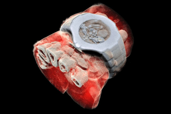
The time has come when black and white X-ray images will replace color three-dimensional pictures of a person's "inner world". For the first time, a New Zealand company that produces color computed tomographs MARS, tested their inventions on humans (they still tried to scan living organisms of a lower order and flora in three-dimensional graphics).
The invention of the new generation Medipix3RX chips allows visualization of the internal structure of a living organism in close proximity to true color perception, volumetric shapes and tissue density. Technologically, the Medipix chip makes it possible to view tissues in a volumetric section, recognizing not only the density of the tissue, but also the nesting of one tissue structure into another. Color X-ray with MARS tomographs is able to reconstruct human tissue with anatomical details.
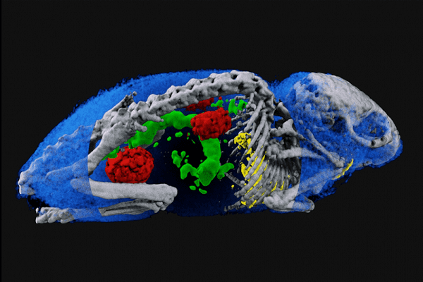
A technological breakthrough in the work of Medipix is based on the chip's ability to measure the length of a radio wave that decays when passing objects of different densities (taking into account the molecular structure), which serves as a source for distinguishing the qualitative characteristics of tissues and radiopaque substances, that is, on ordinary X-rays, bones look white and radiopaque Iodine appears white, but under the new scanning conditions bones appear green and iodine yellow, allowing different tissues to be identified. Even those formations, the structure of which is unknown, will be visually separated from normal tissues.
Chip - detector Medipix3RX distinguishes lipids, water, tissues, pharmaceuticals (including barium, iodine and gadolinium), nanoparticles, that is, low-contrast substances (not visible with conventional X-rays) and high-contrast substances (visible with conventional X-rays). This provides advantages in the study and diagnosis of diseases such as obesity of the liver, obesity of tissues, atherosclerosis of blood vessels, and malignancy of tissues.
Manufacturers of Medipix3RX technology claim that the chip is capable of visualizing the cellular level of tissue structure, which is useful for histological studies without the participation of radioisotopes. In addition to image accuracy, new-generation color X-ray tomographs generate lower radiation doses, which makes radiology safe and creates prerequisites for non-one-time scanning of tissues.

Read

Read


