Ophthalmology >>>> Intraocular pressure treatment
Intraocular pressure treatment.
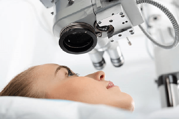
The level of ocular pressure has a significant effect on the condition of the eye and the correct functioning of the optical system, therefore, differences in ocular pressure must be monitored and regulated in time. Intraocular pressure in a normal state does not go beyond the limits of values below 9 mm Hg or above 24 mm Hg.
Reasons for changes in intraocular pressure:
- Inflammatory processes that develop in the tissues of the eye (especially chronically occurring);
- Injuries to the eyeball and oculomotor apparatus;
- The development of tumors in the eye area;
- Hypertension or hypotension;
- Renal failure;
- Diseases of the thyroid gland;
- Dehydration.
Increased eye pressure.
- Since the filling of blood vessels is directly related to the level of intraocular pressure, it is necessary to control the increase in blood pressure and reduce it in time, avoiding a persistent increase in its indicators.
- Avoid eye fatigue, develop a certain regime of rest for the eyes and try to adhere to it daily.
- Treat diseases of a neurological nature so that they do not aggravate the condition of the motor nerves of the eye.
- Control renal pressure in kidney disease (kidney failure, absence of one kidney), drink regularly diuretics and drugs that lower the level of nitrogen in the blood.
- Prevent or treat thyroid disorders.
- Avoid taking alcoholic beverages due to the fact that alcohol has a vasodilating effect (which is especially reflected in the eyes).
As a complication of high intraocular pressure, glaucoma oratrophic changes in the optic nerve can develop .
Symptoms of increased intraocular pressure:
- Headache develops in the forehead or temples
- Painful movements of the eyeball
- Feeling of distention of the eyeball
- Feeling as if the eyes are covered with sand
- Redness of the white of the eye
- Decreased field of view
- Deterioration of vision clarity
- Decreased clarity of twilight vision
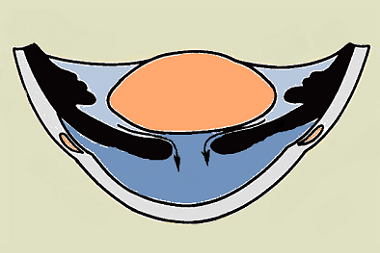
Decreased eye pressure.
For a person, a decrease in intraocular pressure can go unnoticed; it can be detected by examining the eyeball. In severe cases, the eyeballs sink slightly into the orbit.
With a decrease in eye pressure for a long period, visual impairment occurs. A persistent decrease in intraocular pressure will cause the atrophy of the eyeball, its wrinkling (this process is irreversible).
Intraocular pressure treatment methods:
Conservative treatment relies on drugs that lower the pressure of the intraocular fluid:
- Cholinomimetics - improve the outflow of intraocular fluid,
- Alpha-adrenergic agonists - promote the outflow of fluid from the anterior chamber of the eye,
- Beta-blockers - regulate the production of intraocular fluid, reducing its volume,
- Prostaglandins - promote the outflow of intraocular fluid along the posterior path.
Intraocular pressure surgical treatment is carried out in the absence of drug therapy results.
Operations are performed:
- Iridotomy - a micro- hole (one or more) is made by a laser in the iris to drain the eye fluid. The outer shell of the eye is not damaged.
- The outer wall of the scleral sinus is excised (sinusotomy) and the trabecular wall is stretched using laser microcircuits (trabeculospasis).
- Goniotomy - cut through the fusion in the area of the iris-corneal angle of the anterior chamber of the eye.
- Trabeculotomy - a laser is used to cut the trabecular meshwork (the tissue that connects the edge of the iris and the posterior plane of the cornea), which expands the cells of the trabecular meshwork and increases the outflow of ocular fluid.

Read

Read


