Ophthalmology >>>> Intraocular pressure
Intraocular pressure.

Interstitial pressure (the so-called extracellular pressure of interstitial fluid) maintains the turgor of all organs and tissues in the body. Its value does not exceed two / three mm p. with. But there is intraocular pressure (IOP), which in its physiological characteristics exceeds the pressure of the tissue fluid and its indicators fluctuate in the range of 9 - 22 mm p. with.
The constancy of the level of intraocular pressure is the most important factor in maintaining normal eye homeostasis. Intraocular pressure maintains a stable spherical shape of the eye, straightens all the membranes of the eye, regulates the relationship of the internal structures of the eye with each other, is responsible for metabolic processes and removes metabolic products from the eye. Along with these functions, intraocular pressure is responsible for the correct function of the ocular optical system.
The entire process of eye hemodynamics (blood circulation through the blood vessels of the eye) is associated with fluctuations in intraocular pressure. But the hydrodynamic equilibrium of the eye ensures the outflow and inflow of intraocular fluid, which thus maintains approximately the same level of intraocular pressure.
It has been experimentally established that rapid changes in the blood filling of the eye vessels change the intraocular pressure, and slow changes in the blood filling of the eye vessels do not affect the dynamics of the intraocular pressure. With a decrease in the blood filling of the eye vessels, the tone decreases and the rate of outflow of fluid from the eyeball proportionally decreases. In this case, the level of outflow of fluid from the eyeball becomes lower than its inflow into the eyeball, which leads to the restoration of the initial indicators of intraocular pressure. This is a very important mechanism for regulating intraocular tone, since it plays a protective role for the eye in case of deformities of the eyeball during movement or blinking, in case of accidental compression of the eye, with fluctuations in blood pressure,
As noted, the constancy of the level of intraocular pressure is regulated by special mechanisms that ensure the formation of fluid, its outflow and inflow. Aqueous humor ( intraocular fluid ) is produced by the ciliary body. It is believed that the regulator of the secretion of aqueous humor is the hypothalamus. The fluid circulates almost always in the anterior part of the eye. In its composition, it differs from blood plasma: less in density and more acidic, since the concentration of ascorbic acid in it is twenty-five times higher than in blood plasma. In its composition, the intraocular fluid is more similar to the cerebrospinal fluid. The reservoirs of the intraocular fluid are located to a greater extent in the anterior and to a lesser extent in the posterior chambers of the eye.
Abnormal decrease in secretory functions for the production of intraocular fluid lead to a number of diseases: iridocyclitis, hypotension of the eye, subatrophy and atrophy of the eyeball. An excessive increase in the secretion of intraocular fluid leads to a persistent increase in intraocular pressure - ophthalmic hypertension . The term "ophthalmic hypertension" is used in cases where, with two consecutive measurements, IOP exceeds 21 mm p. with. According to a study of a healthy population, the average values are 16 mm Hg with small deviations. In older people, especially women, these indicators are 24 mm p. with. instead of the usual 21 mm p. with.
It has been proven that the movement of intraocular fluid along the drainage system of the eye experiences resistance 100,000 times greater than when blood moves through the vessels. Such a high level of resistance to the outflow of fluid from the eye, plus the slow rate of its secretion, provides just the optimal level of intraocular pressure that is necessary for a healthy body. But with an abnormally obstructed outflow of fluid, a complex disease develops in more than 90% of cases - glaucoma.
The value of intraocular pressure is the indicator that, along with others, is involved in the diagnosis of diseases of hypertension or hypotension of the eye , iridocyclitis, glaucoma and other diseases.
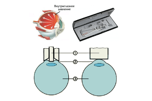
It is very problematic to measure the absolute value of the ocular pressure (its true indicator); for this, a thin cannula connected to a tonometer must be inserted into the eye (this cannot be done in living patients). Therefore, to make diagnoses, the relative value of intraocular pressure is used, which can be measured in two ways: approximate (palpation) and tonometric.
Palpation can determine the density of the eye. In this case, the patient must look down in order to avoid pain or discomfort, the doctor rests on the patient's forehead with his little finger, ring and middle fingers of both hands, and carefully puts both index fingers at a distance from each other on the upper eyelid, above the upper edge of the cartilage, and palpates with one finger the eyeball, and with the other finger, lightly presses on it from the opposite side. The magnitude of the eye pressure is judged by the density of the eyeball, that is, by the compliance of the sclera to pressure: if the intraocular pressure is high, then it takes a lot of effort to flatten the sclera. If intraocular pressure is normalor low, then light tremors are felt with minimal pressure on the sclera. This method is used only when it is not possible to conduct a hardware study of intraocular pressure.
With the tonometric method of measuring intraocular pressure, ophthalmotonometers (impression and applanation) are used. In the applanation method, the patient is placed on a couch, the eyes are instilled with a local anesthetic, forced to look forward, the eyelids are spread, and a special load painted with dye is lowered onto the cornea. Under the influence of the load, the cornea is flattened, the area of contact with the load increases, the dye on the load is washed off by a tear, and an unpainted circle remains on the load, the diameter of which is measured and the indicators are applied to a special grid of coordinates. Changing the weight of the goods, the data are consistently applied and the results are analyzed. The impression method differs in that instead of a weight, pins (3 mm in diameter) are used, which depress the membranes of the eye and form pits.
Today, there are tonometric electronic devices that use platforms instead of weights, connected to a device (sensor) that fixes the degree of compression. There is also a non-contact method for determining intraocular pressure using a jet of compressed air. This does not require local anesthesia, there is no risk of infection, but the measurement accuracy is reduced.

Read

Read


