Neurological diseases >>>> The principles of the vestibular analyzer
The principles of the vestibular analyzer.
The vestibular apparatus is a highly complex sensory system that keeps the body from falling in three-dimensional space. The work of the vestibular apparatus is carried out using a vestibular analyzer. Its main function is to organize balance by analyzing the body's presence in space and its movement (acceleration; deceleration; overturn; movement up, down, sideways). To be precise, the analyzer monitors the position and movement of a person's head. In order to understand whether your vestibular analyzer is functioning correctly, what are the prerequisites for disturbances in its work, you need to have an idea of its structure and interaction with different systems and organs.
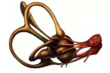
It's no secret that the vestibular apparatus is located in the middle ear, but where exactly? The inner ear of a person consists of a bony labyrinth and a membranous labyrinth (membrane) included in it, repeating the configuration of the bony labyrinth. The bony labyrinth is located deep in the temporal bone pyramid. This bone formation has three sections: if you mentally draw a trajectory in the direction starting from the auricle to the middle ear and further into the depth of the cranium, then first there is the cochlea, then the vestibule, then a system of three semicircular canals. The channels are located in mutually perpendicular three planes. Membranous semicircular canals (located inside the corresponding bone structure of the semicircular canals) are filled with a special fluid – endolymph which has a positive electrical charge.
The vestibule (or middle section) has two pockets: spherical and elliptical. The spherical pocket is located near the cochlea and has a spherical pouch inside, the elliptical pocket is adjacent to the semicircular canals and has an elliptical pouch inside.
Vestibular sensory cells are located in five receptor regions: in three semicircular canals and in the two vestibular sacs described above. Special peripheral fibers (axons) lead to the hairs of these receptor cells from the vestibular node that runs in the internal auditory canal (called the “vestibular ganglion”). Other, so-called central fibers (dendrites) are part of the seventh pair of cranial nerves and lead to the nuclei in the medulla oblongata.
When the endolymph moves in one of the three planes, the hairs of the sensory cells bend, transmitting a signal along the nerve fibers in the appropriate direction. The movement in any of the three planes of the endolymph reflects the movement of the human body in the same plane (or rather, his head).
Considering the structure of the neural connections of the vestibular analyzer from the standpoint of the clinic and diagnosis of diseases of this apparatus, it is necessary to note five connections of the analyzer with various systems and organs.
- Vestibulospinal connections - provide the connection of the analyzer receptors with the muscle system.
- Vestibulo-oculomotor connections - provide a connection with the oculomotor muscles.
- Vestibulovegetative connections - lead to the vagus nerve, diencephalic region (hypothalamus - pituitary region).
- Vestibulocerebellar connections - control the cerebellum.
- Vestibulocortical connections - directed to the temporal lobe of the cerebral cortex.
These five connections correspond to five reactions of the vestibular apparatus:
- Vestibulosomatic reactions distribute muscle tone while accelerating the body;
- Oculomotor reactions are associated with the occurrence of nystagmus - involuntary movements of the eyeballs with slow or fast movement, it is easier to say during nystagmus, the eyes are photographed with the eyes of the picture leaving when the human body moves (fast, slow, rotational);
- Vestibulo-vegetative reactions adapt the body to changes, they are manifested in the form of an increase in blood pressure, an increase in heart rate, the occurrence of nausea and the urge to vomit when accelerating ;
- Vestibulocerebellar reactions redistribute muscle tone , thereby supporting the body in space during movement and acceleration;
- Vestibulocortical connections with the cerebral cortex are responsible for vertigo .
The vestibular analyzer is a highly sensitive mechanism, it reacts sensitively to any changes occurring in the body, especially pathological ones, for example, a brain tumor, even being at a considerable distance from the vestibular apparatus, disrupts its work. The vestibular apparatus has extensive anatomical and physiological connections with various organs and systems and, accordingly, all malfunctions in these organs and systems are adversely reflected in the work of itself.
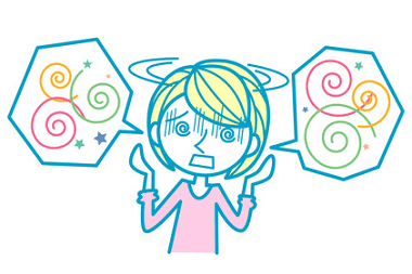
If symptoms such as impaired coordination , dizziness, nausea occur when stretching the neck, turning the head in different directions, or the manifestation of these symptoms without reference to any movement, one can suspect violations either in the work of the vestibular apparatus itself, or pathological disorders of organs and their systems, "Noticed" by the vestibular analyzer, therefore it is necessary to carry out examinations in a timely manner and identify the causes of the unhealthy reaction of the vestibular analyzer.
Such a state of the body as kinetosis (motion sickness) is not considered a pathology, but rather a reaction of an untrained vestibular apparatus to unusual movements and changes in body position in space. But the vestibular analyzer, with a continuous and long-term change in the position of the body (head), is able to adapt to such changes. Therefore, if there are no organic disorders of the vestibular apparatus, it can be trained.


Read
Read