Ophthalmology >>>> Optic nerve atrophy
Optic nerve atrophy.
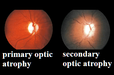
With atrophy of the optic nerve, a decrease in the diameter of the optic nerve is considered characteristic due to a decrease in the number of axons (processes of the nerve cell). In this case, blanching of the optic nerve head develops and visual functions are impaired: defects of the visual field appear, the degree of visual acuity decreases. Visual acuity can vary from 0.7 and, practically, to blindness. There are primary atrophic changes in the optic nerve (as a result of compression compression by a tumor or aneurysm, as a consequence of retrobulbar neuritis, optic neuropathies, toxic or other effects) or secondary. Secondary atrophic changes in the optic nerve are preceded by edema of the optic nerve head, chronic inflammation, optic neuropathy, papillitis. Optic atrophy can be congenital or acquired.
Acquired atrophy develops against the background of injury to the optic nerve fibers or retinal cells. In this case, fibers are damaged at different levels: at the level of the orbit, the optic canal, and the cranial cavity.
Causes of optic nerve atrophy (acquired):
- injury
- inflammation
- high level of intraocular pressure
- violation of the vascular circulation that feeds the optic nerve, with glaucoma and other diseases
- toxic damage
- metabolic disorders
- compression of nerve fibers by a tumor
- degenerative processes in the eyeball area
- myopia and others.
Blanching of the optic nerve can occur gradually over time, the degree of blanching depends on the remoteness of the lesion, on the nature of the disease that caused disturbances in the structure of the optic nerve. When the cause of the disorders is a tumor, then clinical manifestations can manifest themselves only in the form of visual disturbances, and changes in the fundus appear within a few weeks or months.
If the cause of damage to the optic nerve is trauma or inflammation, then the first signs of atrophy appear after which number of days (or weeks).
Atrophic changes in the optic nerve can be primary (as a result of compression compression by a tumor or aneurysm, as a consequence of retrobulbar neuritis, optic neuropathies, toxic or other effects) or secondary. Secondary atrophic changes are preceded by edema of the optic nerve head, chronic inflammation, optic neuropathy, papillitis.
Congenital atrophic changes in the optic nerve are of a genetic nature. They occur in two forms: autosomal dominant, in which visual acuity decreases asymmetrically from 0.8 to 0.1; or autosomal recessive, when vision drops to almost blindness, starting in childhood.
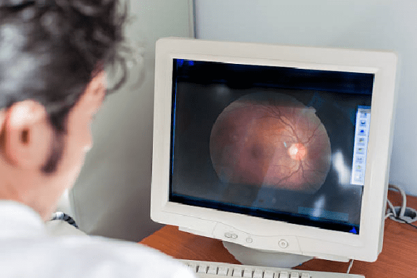
For diagnostic measures to identify atrophic changes in the optic nerve:
- measure intraocular pressure,
- determine visual acuity,
- reveal the boundaries of the field of view in green, white and red colors,
- examine the eye sockets and the state of the brain using MRI.
Treatment of optic nerve atrophy includes symptomatic therapy and treatment of diseases that cause atrophic changes in the optic nerve.
Symptomatic therapy includes:
- vitamin therapy (C and group B),
- drugs: improving metabolic processes in tissues, relieving inflammation, improving blood supply to the vessels approaching the optic nerve,
- stimulating therapy (electro-, magnetic or laser stimulation of the optic nerve).


Read
Read