
Endocrinology >>>> Paget bone disease
Paget bone disease.
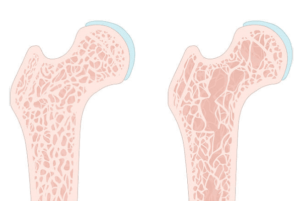
Paget disease (synonyms: osteodystrophy, osteitis deformans) is a chronically current disease of bone tissue, when the process of bone modeling is disrupted and foci of increased tissue resorption appear, which are replaced with time by defective bone tissue. This disruption of bone remodeling changes the structure of the skeleton, causing outwardly noticeable bone deformities and fractures.
The causes of Paget disease lie in the appearance of abnormally large osteoclasts and are associated with genetic characteristics and, possibly, viral infections. The origin of the disease remains unclear.
Signs of Paget disease:
- Bone pain (including in the area of the vertebrae),
- In case of damage to the bones of the skull, headaches may be disturbed,
- Above the site of bone deformation, painful swelling, reddening of the skin and possibly an increase in the temperature of certain tissue areas occur,
- Bone deformation,
- There is osteoarthritis of the joints,
- Frequent bone fractures,
- Neurological complications in the form of deafness, which are caused by overgrown areas of the bones of the skull, compressing nerve fibers.
Paget disease can be diagnosed by conducting a biochemical analysis to determine the level of alkaline phosphatase, which, in the event of the development of the disease, increases its activity. Further, the diagnosis is confirmed by X-ray or by scintigraphy of the bones of the skeleton (scanning the skeleton using technetium-labeled phosphonates).
It is necessary to differentiate Paget disease from conditions such as bone metastases, hyperparathyroidism, gout, arthritis, osteomalacia, and acromegaly.
Treatment of Paget disease consists of several directions:
- Long-term drug therapy with first-line antiresorptive drugs (nitrogen-containing biophosphonates, Risedronic acid, Alendronic acid, Pamidronic acid, Zolendronic acid), second-line (calcitonin), analgesics as needed.
- Surgical treatment (including endoprosthetics) for fractures and bone deformities.
- Wearing orthoses that relieve extremities during an exacerbation of the disease.
When remission is achieved, characterized by a decrease in the intensity of pain, the absence of new foci of deformity, normalization of the activity of alkaline phosphatase, X-ray control is carried out every three years and the state of alkaline phosphatase is checked every six months.


Read
Read