Emergencies >>>> What to do in case of bleeding in the eye?
What to do in case of bleeding in the eye?

The causes of hemorrhage in the eye can be trauma or the development of pathological conditions in the structure and functions of the eye. In both situations, the integrity of the blood vessels penetrating the tissues of the eye is violated, the blood goes beyond the vascular wall and enters the tissue adjacent to the rupture site.
The most common eye injuries are:
- Contusion (blunt trauma to the eye) occurs when the eyeball and / or the tissues surrounding the eye are compressed (for example, when struck directly in the eye or on the head). Moreover, an outwardly insignificant injury can lead to significant damage to the eyeball and the surrounding oculomotor apparatus.
- Mechanical trauma to the eye, directly violating the integrity of the eyeball and surrounding tissues (resulting from the use of blunt, stabbing or cutting objects).
Pathological conditions affecting the state of the vascular wall and provoking hemorrhage in the eye:
- Violation of blood circulation as a result of atherosclerosis or thrombosis of the blood vessels of the eye, narrowing the lumen of the vessels;
- Tumors that compress and damage blood vessels;
- Aneurysms of the vascular wall;
- Changes in the structure of the vascular wall itself (reduced elasticity, fragility, increased permeability) and its tendency to cracks, ruptures;
- Abnormal development of the eye blood supply system;
- High blood pressure;
- Increased intraocular pressure;
- Inflammatory processes that disrupt the condition of the tissues of the eye, including blood vessels;
- Physical stress when lifting weights, coughing, pushing.
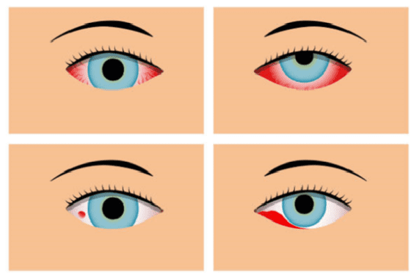
What types of bleeding in the eye can occur:
- Into the eye socket,
- Into the anterior chamber of the eye,
- For the conjunctiva of the eye,
- Into the retina of the eye,
- Into the vitreous body.
Hemorrhage into the orbit may not be noticeable on a superficial examination, but its existence is suggested based on some signs:
- The eyeball is displaced outward from the orbit and "bulging" appears,
- Limited movement of the eyeball appears,
- Hemorrhage is located in the thickness of the skin of the eyelids or conjunctiva.
Hemorrhage in the orbit is most often found in trauma to the skull, brain, eye contusions, sometimes in the case of certain diseases (violations of the physical characteristics of the vascular wall, vasculitis, aneurysms).
If the described signs of bleeding in the orbit appear, it is necessary to urgently consult a doctor, since the damage can be life-threatening.
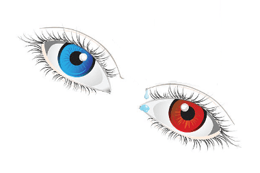
Hemorrhage into the anterior chamber of the eye or hyphema looks like a homogeneous formation of red color, which changes its contours depending on the position (horizontal or vertical) of the person. In the vertical position, it flows down (to the bottom of the anterior chamber of the eye); in the horizontal position, the blood spreads evenly throughout the entire space of the anterior chamber. If the hyphema does not cover the pupil area, then it does not interfere with vision. Hyphema usually resolves within a few days, but if this does not happen within 10 days, then this indicates the development of a pathological condition (uveitis, cataracts, glaucoma) in the eye area and requires the intervention of an ophthalmologist.
Subconjunctival bleeding or hyposhagmus occurs under the conjunctiva in the area between the conjunctiva and the sclera of the eye. The cause of this hemorrhage is the rupture of small vessels that penetrate the conjunctiva. Subconjunctival hemorrhage often occurs as a result of high blood pressure, exertion, physical exertion, antiplatelet or fibrinolytic drugs.
The hyposhagmus is not life-threatening and resolves within one to three weeks like a bruise. The bloody spot behind the conjunctiva is usually limited, but if the borders of the spot expand, then this indicates that the bleeding continues, and in this case, an urgent need to see a doctor.
Retinal hemorrhage is characterized by several typical signs that disrupt the functioning of the visual apparatus:
- Blurring the edges of the image,
- Decreased vision,
- Flickering flies or a mesh in front of the eyes, moving with the movement of the eyes.
Treatment for this type of hemorrhage depends on the extent to which the retina is involved in the bleeding process. With extensive hemorrhages in the retina, urgent hospitalization of the victim is indicated. The severity of bleeding into the retina can only be determined by a doctor, therefore, in the presence of the above symptoms, it is necessary to consult a specialist.
Hemorrhage into the vitreous body or hemophthalmus occurs as a result of serious damage to the vessels of the eye and requires the immediate assistance of an ophthalmologist, since it is dangerous with loss of vision. Untimely intervention can lead to complications: atrophy of the eyeball, retinal rupture, retinal detachment, hemolytic glaucoma.
Hemophthalmos is characterized by the appearance of dark mobile spots before the eyes and / or flashes of light. When hemophthalmos is detected, the patient is recommended to sleep in the "half-sitting" position so that the blood settles under the influence of gravity and gives access to the fundus examination.
With small hemorrhages in the vitreous body, resorption occurs independently without special treatment.
If you notice any of the above signs of hemorrhage, the first step is to stop using blood thinners (if you are using them, of course). These drugs include aspirin and drugs containing acetylsalicylic acid, heparin, lyoton, vitamin K antagonists and other similar drugs. These drugs hinder the processes of thrombus formation and will not allow bleeding to stop.
In the complex cases described above, with bleeding into the eye, immediate help from an ophthalmologist is required. Do not delay consulting an ophthalmologist, otherwise you may lose your eyesight.


Read
Read